BC Doctors of Optometry

The Overlooked Predictor in Cataract Co-Management

Brought to you by Meivertor
Why upper eyelid meibography matters for Canadian ODs and their post‑op patients


Cataract surgery delivers consistently strong visual outcomes. Patients expect clearer vision, sharper reading, and a smooth recovery. What many do not expect—and what their co-managing optometrist is often the first to hear about—is the dry eye that can follow.

Between 37% and 55% of patients develop dry eye symptoms after cataract surgery, including some who had no pre-existing complaints. ¹,² For some, symptoms are short-lived. For others, burning, fluctuating vision, and foreign body sensation persist for weeks to months, diminishing an otherwise excellent surgical result.

For Canadian ODs involved in cataract co-management, that matters twice. These are often the patients who return first with clouding, irritation, or complaints that “the surgery went fine, but the vision still doesn’t feel right.” And in systems where patients may wait months between referral and surgery, there is often more opportunity than many practices realize to identify risk factors early, clean up the ocular surface, and set expectations before the surgical date arrives.

As optometrists take on a larger role in perioperative care, attention is shifting earlier in the process. Identifying who is at risk before surgery—not after—can change the course of the post-op experience.

Emerging evidence points to a specific, and often overlooked, predictor: the meibomian glands of the upper eyelid.

Why post-cataract dry eye happens

Cataract surgery places temporary stress on the ocular surface through several well-recognized factors. The surgical environment itself, preoperative prep, and postoperative medications all contribute to disruption of the tear film and ocular surface stability. ³,⁴

During and after surgery, corneal sensitivity is reduced, tear film dynamics are altered, and the ocular surface is exposed to topical medications used multiple times daily for weeks. These factors can destabilize the tear film, particularly in patients with underlying gland dysfunction. ³,⁴

When meibomian gland function is already compromised, the ocular surface is less able to compensate. The lipid layer becomes less stable, tear breakup time shortens, and evaporative dry eye establishes more quickly. ⁴ In patients considering premium or Toric IOLs, that instability may also affect preoperative measurements, visual consistency, and overall satisfaction with the surgical experience.

Surgery does not cause dry eye. More often, it exposes an underlying vulnerability that was already present. ³,⁴

What the data show about the upper lid

Fujimoto and colleagues (Clinical Ophthalmology, 2022) examined 82 eyes of 43 patients who had no subjective dry eye symptoms before uncomplicated cataract surgery. One month postoperatively, 25.6% had developed new-onset symptoms. ⁵

Multivariable logistic regression identified two statistically significant independent predictors: preoperative upper eyelid meibomian gland loss area (odds ratio 6.72; p = 0.012) and female sex (odds ratio 4.20; p = 0.037). ⁵ Lower eyelid gland loss was not a significant predictor in this cohort.

That last point carries the most clinical weight. A patient with reassuring lower lid meibography and significant upper lid dropout will not be flagged by a lower-lid-only protocol. But according to these findings, that patient is the one at highest risk.

“A comprehensive meibography assessment should always include both upper and lower eyelids. Relying solely on lower eyelid imaging risks missing early gland changes that could guide timely intervention and improve patient care.”
 Paul M. Karpecki, OD,  Review of Optometry, May 2025

Why the upper lid is structurally more vulnerable

The upper eyelid contains more meibomian glands than the lower, and those glands are longer and more anatomically slender. That structure makes them susceptible to early morphological changes—tortuosity, thickening, partial dropout—that can precede both symptoms and lower-lid findings. ⁶

Gupta and Karpecki’s 2025 review in Cornea synthesized nearly two decades of meibography data and concluded that upper eyelid changes often appear before equivalent changes in the lower lid—and that the status of the two lids does not reliably predict each other. Lower lid findings cannot be used as a proxy for the upper.

Because the upper eyelid makes primary contact with the ocular surface on each blink, upper lid gland compromise can have an outsized effect on tear film stability relative to gland count alone. In practical terms, every blink becomes a stress test the tear film may no longer pass.


Clinical implications for pre-surgical assessment

For ODs co-managing cataract patients, the preoperative evaluation can be the point of greatest impact. Risk factors for postoperative dry eye are often already present at the time of referral, making it important to identify them early rather than after symptoms develop.

Incorporating bilateral meibography—with careful attention to the upper lid—into the pre-referral workup provides practical value: it helps stratify risk, supports a rationale for initiating treatment before surgery, and establishes a baseline for more meaningful postoperative comparison.

That is especially relevant when there is a long interval between referral and the actual surgical date. Instead of viewing wait time as passive delay, practices can use it as active preparation time: identify gland loss, begin a dry eye clean-up program, improve tear film quality, and communicate clearly with the surgeon about what has already been found and treated. 

For patients, that preparation can support a better overall experience. For the practice, it can mean cleaner measurements, better informed IOL discussions, fewer postoperative surprises, and a more confident handoff back and forth between optometrist and surgeon.

A practical conversation to have before referral

One of the most valuable uses of upper lid meibography is not just diagnostic—it is conversational. When patients can see that gland loss or dropout is already present before surgery, it becomes much easier to explain why preoperative treatment matters and why perfect surgery does not always guarantee a symptom-free recovery.

A simple chairside explanation may sound like this:

“Your cataract surgery can still go very well, but these images show that the oil glands in your eyelids are already under some stress. That means you may be more likely to notice dryness, fluctuating vision, or irritation afterward—even if the surgery itself is perfect. The good news is we can get ahead of that now. We have time to clean up the surface, improve the tear film, and give your surgeon better information before the procedure.”

That kind of conversation does three things at once: it builds awareness, sets realistic expectations, and positions preoperative dry eye treatment as part of good surgical planning rather than an added extra.


Pre- and Post-Operative Management Framework

TREATMENT

✓  Lid hygiene and warm compresses

✓  Consider in-office therapies (IPL, thermal pulsation)

✓  Optimize ocular surface before surgical clearance

The full picture

Post -cataract dry eye is not a complication that appears without warning. The warning is often present preoperatively—in the upper eyelid meibomian glands, in a patient population that has historically not had those glands imaged routinely.
As Dr. Cory Lappin observed in Optometric Physician, when it comes to a complete dry eye evaluation, the standard should be to image both the upper and lower glands—or you end up with only half the story.⁷ For cataract co-management, that “other half” may be the part that tells you most about who is likely to struggle after surgery.

  “The key to reliable imaging is consistent lid eversions. Meivertor produces consistent, reproducible lid eversions — and techs love it because it takes very little practice to master.”
Dr. Cory Lappin, OD, MS, FAAO Optometric Physician, Nov 2025

Clinical Tool Note

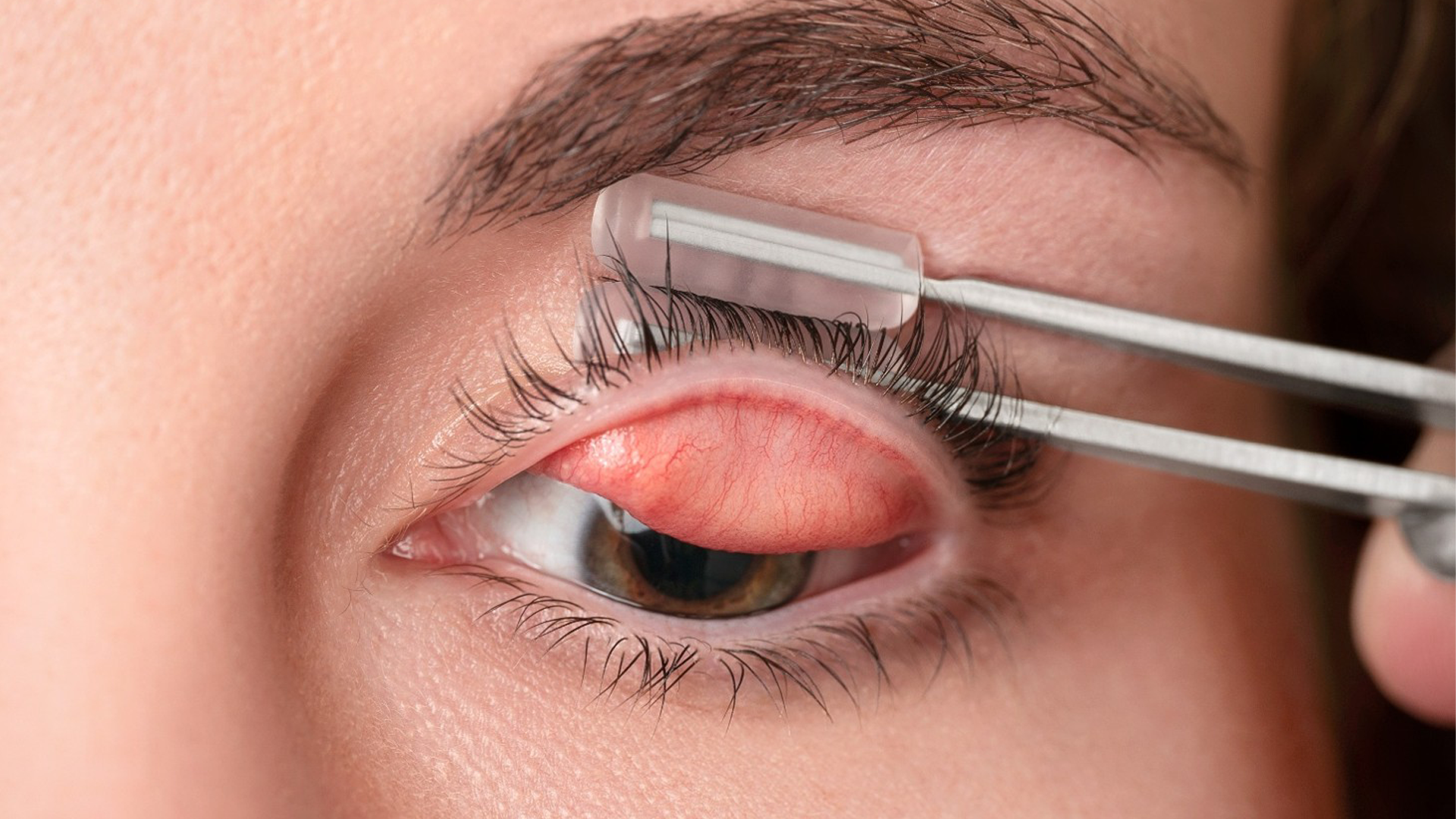
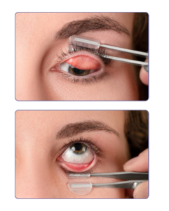
A practical barrier to routine upper lid imaging has been the need for two hands to evert the lid while a third operates the camera.

Meivertor® is a single-handed eyelid eversion instrument with disposable silicone tips that secures the lashes and everts the lid with a controlled rotation—freeing the other hand for the imaging system.

Key features:

  • Single-handed operation
  • Disposable tips (infection control, patient comfort)
  • Consistent, reproducible eversions
  • Minimal technician training required

The tool is easy to use and Meivertor provides excellent staff support to help educate users: How to Use Meivertor

Available in the U.S., Canada, and select international markets. www.meivertor.com

References
1. Miura M, Inomata T, Nakamura M, et al. Prevalence and characteristics of dry eye disease after cataract surgery: a systematic review and meta-analysis. Ophthalmol Ther. 2022;11(4):1309–1332.
2. Kasetsuwan N, Satitpitakul V, Changul T, Jariyakosol S. Incidence and pattern of dry eye after cataract surgery. PLoS One. 2013;8(11):e78657.
3. Moshirfar M, et al. Postoperative Dry Eye Disease Following Cataract Surgery: A Review. Clin Ophthalmol. 2017.
4. TFOS DEWS II Report. Ocular Surface Society. 2017.
5. Fujimoto H, Ochi S, Inoue Y, Kiryu J. Upper eyelid meibomian gland dysfunction is a risk factor for dry eye symptoms following cataract surgery in the early phase. Clin Ophthalmol. 2022;16:293–302.
6. Gupta PK, Karpecki P. Comprehensive assessment of the meibomian glands by meibography: why the upper eyelids matter. Cornea. 2025;44(1):128–135.
7. Lappin CJ. Off the cuff: only half the story. Optometric Physician. November 24, 2025.

Scroll to Top